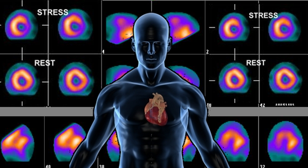
 A nuclear heart scan is a test that provides important information about the health of your heart.
For this test, a safe, radioactive substance called a tracer is injected into your bloodstream through a vein. The tracer travels to your heart and releases energy. Special cameras outside of your body detect the energy and use it to create pictures of your heart.
Nuclear heart scans are used for three main purposes:
To check how blood is flowing to the heart muscle. If part of the heart muscle isn't getting blood, it may be a sign of coronary heart disease (CHD). CHD can lead to chest pain called angina (an-JI-nuh or AN-juh-nuh), a heart attack, and other heart problems. When a nuclear heart scan is done for this purpose, it's called myocardial perfusion scanning.
To look for damaged heart muscle. Damage might be the result of a previous heart attack, injury, infection, or medicine. When a nuclear heart scan is done for this purpose, it's called myocardial viability testing.
To see how well your heart pumps blood to your body. When a nuclear heart scan is done for this purpose, it's called ventricular function scanning.
Usually, two sets of pictures are taken during a nuclear heart scan. The first set is taken right after a stress test, while your heart is beating fast.
During a stress test, you exercise to make your heart work hard and beat fast. If you can't exercise, you might be given medicine to increase your heart rate. This is called a pharmacological (FAR-ma-ko-LOJ-ih-kal) stress test.
The second set of pictures is taken later, while your heart is at rest and beating at a normal rate..